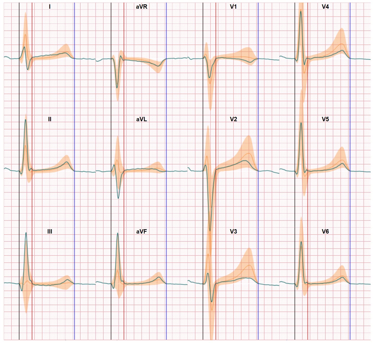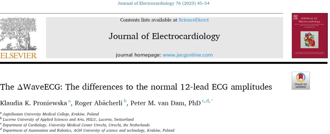
As of today our new Delta map ECG view on 12-lead ECG waveforms is supported by a great article published in the 76 edition of the Journal of Electrocardiology. ECG waveform interpretation is still (111 years after the ECG introduction) a major challenge for physicians. And the Delta map view has been developed to make ECG waveform interpretation easier and better. The ΔWaveECG can distinguish the abnormal from normal ECG waveform segments, making the ECG easier to classify as normal or abnormal. Conduction disorders and ST changes due to ischemia and abnormal T-waves are effortless to detect, also by non-ECG expert readers, thus improving the early detection of cardiac patients. This new view on the ECG might become an important tool in for instance the emergency setting and in referring ECG recording centers with limited experienced.
As such the ΔWaveECG maps could be used to limit the referral of patients with an normal ECG from primary care to next level of healthcare. The examples on which the ΔWaveECG was tested show that conduction disorders shown by QRS deviations, myocardial infarction shown by deviating ST elevation or depression, and abnormal T wave morphology can be easily detected.
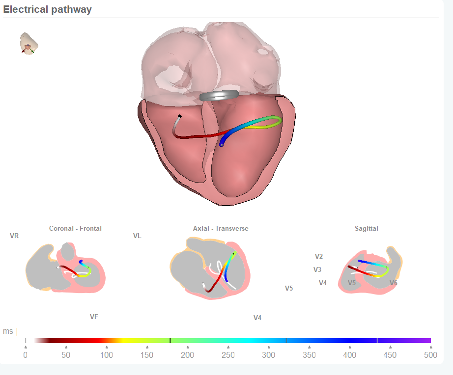
The proposed ΔWaveECG classification, based on the new Delta map ECG view, is different from anything currently developed for the standard 12‑lead resting ECG (to the best of our knowledge). Normal values have been collected for many people, but this did not take into account the whole ECG waveform, only key amplitudes at certain fiducial moments. This also limits the influence of the variability in fiducials by different ECG machine manufacturers. The ΔWaveECG maps compare the whole ST segment to normal values, and thus the exact fiducial values are of no importance. Other methods determine the beat-to-beat variation in an ECG of a patient to detect unstable functional myocardium (ischemia) but this requires ECG recordings much longer than the normal 10 s of the resting ECG. The new delta map ECG view will be incorporated in the CineECG analysis and made available for further testing.

A big thank you to Klaudia Proniewska and Roger Abächerli for their contribution to this study and its results. And to the Dutch Heart Foundation and the Polish National Centre for Research and Development for their support of this research.