
Expertise, experience and the drive behind CineECG
The company behind CineECG
ECG Excellence
They take pride in following the footsteps of Professor Willem Einthoven who co-invented the ECG in 1911. Improving ECG interpretation performance is one the key drivers for the ECG Excellence team to move the classic ECG from the analogue to the digital age. CineECG is the first of a range of ECG improvement technologies the team is working on.
Scientific base for CineECG
A list of relevant CIneECG related publications is shown below
The normaal ventricular CineECG
van Dam PM, Boonstra M, Locati ET, Loh P. The relation of 12 lead ECG to the cardiac anatomy: The normal CineECG. Journal of Electrocardiology. 2021;69:67-74. doi: https://doi.org/10.1016/j.jelectrocard.2021.07.014
In this study we used 6525 12‑lead ECG tracings labelled as normal obtained from the certified Physionet PTB XL Diagnostic ECG Database to construct the CineECG. All 12 lead ECGs were analyzed, and then divided by age groups (18–29,30-39,40-49,50-59,60-69,70–100 years) and by gender (male/female). For each ECG, we computed the CineECG within a generic 3D heart/torso model. Based on these CineECG’s, the average normal cardiac location and direction for QRS, STpeak, and TpeakTend segments were determined. The CineECG direction for the QRS segment showed large variation towards the left free wall, whereas the STT segments were homogeneously directed towards the septal /apical region. The differences in the CineECG location for the QRS, STpeak, and peak-Tend between the age and gender groups were relatively small (maximally 10mmat end T-wave), although between the gender groups minor differences were found in the 4 chamber direction angles (QRS 4°, STpeak 5°, and TpeakTend 8°) and LAO (QRS 1°, STpeak 13°, and TpeakTend 30°). CineECG demonstrated to be a feasible and pragmatic solution for ECG waveform interpretation, relating the ECG directly to the cardiac anatomy. The variations in depolarization and repolarization CineECG were small within this group of normal healthy controls, both in cardiac location as well as in direction.
The normal atrial CineECG
Locati ET, Pappone C, Heilbron F, van Dam PM. CineECG provides a novel anatomical view on the normal atrial P-wave. European Heart Journal – Digital Health. 2022;3:169-180. doi: 10.1093/ehjdh/ztac007
From 6409 normal ECGs from PTB-XL database, we computed a median beat with fiducial points for P-and Q onset. To determine the temporo-spatial location of atrial activity during PQ-interval, CineECG was computed on a normal 58-year-old male atrial/torso model. CineECG was projected to three major cardiac axes: posterior anterior, right-left, base-roof, and to the standard cardiac four-chamber, left anterior oblique, and right anterior oblique (RAO) views.
In 6409 normal subjects, during P-wave, CineECG moved homogeneously from right atrial roof towards left atrial base (-54 ± 14 in four-chamber view, 95 ± 24 RAO view). During terminal PQ-interval, the CineECG direction was opposite, moving towards left atrial roof (62 ± 27in four-chamber view, 78 ± 27 RAO view). We identified the deflection point, where the atrial CineECG changes in direction. The time from P-onset to deflection point was similar to P-wave duration. CineECG provided a novel three-dimensional visualization of atrial electrical activity during the PQ-interval, relating atrial electrical activity to the atrial anatomy. CineECG location during P-wave and terminal PQ-interval were homogeneous within normal controls.
Normal ECG waveforms
Proniewska KK, Abächerli R, van Dam PM. The ΔWaveECG: The differences to the normal 12‑lead ECG amplitudes. Journal of Electrocardiology. 2023;76:45-54. doi: https://doi.org/10.1016/j.jelectrocard.2022.10.014
Creation of a normal ECG amplitude distribution to enable the distinction by non-ECG experts of normal from abnormal waveforms of the standard 12 lead ECG.
The ECGs of healthy normal controls in the PTB-XL database were used to construct a normal amplitude distribution of the 12 lead ECG for males and females. All ECGs were resampled to have the same number of samples to enable the classification of an individual ECG as either normal or abnormal, i.e. within the normal amplitude distribution or outside, the ΔWaveECG.
From the same PTB-XL database six ECG’s were selected, normal, left and right bundle branch block, and three with a myocardial infarction. The normal ECG was obviously within the normal distribution, and all other five showed clear abnormal ECG amplitudes outside the normal distribution in any of the ECG segments (QRS, ST segment and remaining STT segment).
The ΔWaveECG can distinguish the abnormal from normal ECG waveform segments, making the ECG easier to classify as normal or abnormal. Conduction disorders and ST changes due to ischemia and abnormal T-waves are effortless to detect, also by non-ECG expert readers, thus improving the early detection of cardiac patients.
CineECG in conduction disorders
Boonstra MJ, Hilderink BN, Locati ET, Asselbergs FW, Loh P, van Dam PM. Novel CineECG enables anatomical 3D localization and classification of bundle branch blocks. EP Europace. 2021;23:i80-i87. doi: 10.1093/europace/euaa396
Five hundred 12-lead ECGs were used from the online Physionet-XL-PTB-Diagnostic ECG Database with a certified ECG diagnosis. The mean temporo-spatial isochrone trajectory was calculated and projected into the anatomical 3D heart model. We established five CineECG classes: ‘Normal’, ‘iRBBB’, ‘RBBB’, ‘LBBB’, and ‘Undetermined’, to which each tracing was allocated. We determined the accuracy of CineECG classification with the gold standard diagnosis. A total of 391 ECGs were analyzed (9 ECGs were excluded for noise) and 240/266 were correctly classified as ‘normal’, 14/17 as ‘iRBBB’, 55/55 as ‘RBBB’, 51/51 as ‘LBBB’, and 31 as ‘undetermined’. The terminal mean temporal spatial isochrone contained most information about the BBB localization. CineECG provided the anatomical localization of different BBBs and accurately differentiated between normal, LBBB and RBBB, and iRBBB.
CineECG in progressive AV conduction disturbances
van der Schaaf I, Kloosterman M, Boonstra MJ, van Dam PM, Gorgels APM. CineECG illustrating the ventricular activation sequence in progressive AV-junctional conduction block. Journal of Electrocardiology. 2023;78:1-4. doi: https://doi.org/10.1016/j.jelectrocard.2023.01.004
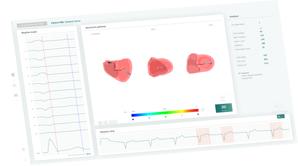
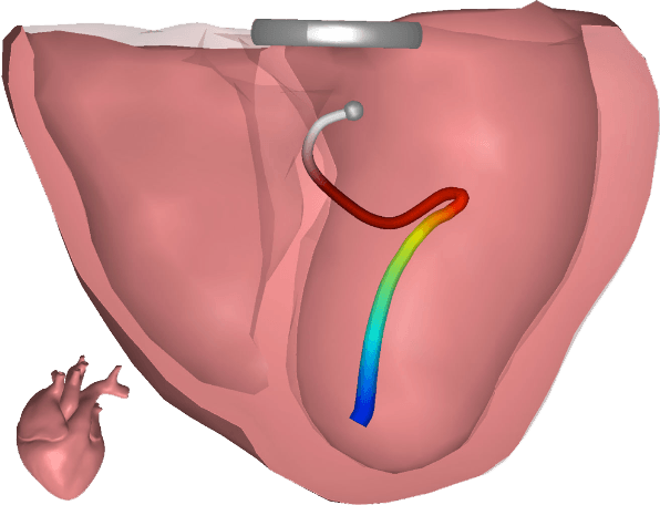
CineECG is used to visualize abnormal ventricular activation in a case of a complex conduction disorder. CineECG combines the standard 12‑lead surface ECG with a 3D anatomical model of the heart. It projects the location and direction of the average ventricular activation and recovery on the heart model over time. In this case, CineECG was able to visualize the different type of fascicular conduction in this progressive conduction block. This novel imaging technique was able to provide additional insight in this complex case, and might be of use in other complex ECG patterns.
CineECG in COVID patients
van Dam PM, Boonstra M, Roudijk R, Linschoten MP, Locati ET, Ciconte G, Borrelli V, Santinelli V, Vicedomini G, Monasky M, et al. The Electro-Anatomical Pathway for Normal and Abnormal ECGs in COVID Patients. Paper/Poster presented at: 2020 Computing in Cardiology; 13-16 Sept. 2020, 2020;
The ECGs of 100 normal controls were used to obtain the normal CineECG paths values for the QRS, ST segment and T-wave. These normal CineECG values were used to classify the COVID-19 ECGs as either as normal or abnormal of 107 patients being treated for COVID-19 in the University Medical Center Utrecht. The CineECG was able to classify 98% of the normal ECG correctly and 94% of the abnormal ECG in comparison to expert ECG classifications. The ability of the CineECG to relate the ECG to the cardiac anatomy supports the detection of abnormal ECGs.
CineECG in Brugada patients
Dam PMv, Locati ET, Ciconte G, Borrelli V, Heilbron F, Santinelli V, Vicedomini G, Monasky MM, Micaglio E, Giannelli L, et al. Novel CineECG Derived From Standard 12-Lead ECG Enables Right Ventricle Outflow Tract Localization of Electrical Substrate in Patients With Brugada Syndrome. Circulation: Arrhythmia and Electrophysiology. 2020;13:e008524. doi: doi:10.1161/CIRCEP.120.008524
In spontaneous and Ajmaline-induced BrS patients, the CineECG localized the late QRS activity in the RVOT, a phenomenon never observed in normal, RBBB, or Ajmaline-negative patients.
CineECG in ST depression patients
Frosted R, Paludan-Muller C, Vad OB, Olesen MS, Bundgaard H, van Dam P, Christensen AH. CineECG analysis provides new insights into Familial ST-segment Depression Syndrome. Europace. 2023. doi: 10.1093/europace/euad116
Familial ST-segment Depression Syndrome (Fam-STD) is a novel inherited cardiac disease associated with arrhythmias and sudden cardiac death. This study was aimed at investigating the cardiac activation pathway in patients with Fam-STD, modelling the electrocardiogram (ECG) phenotype, and performing in-depth ST-segment analyses.
High-resolution ST-segment analyses were performed per lead by dividing the ST-segment into nine 10 ms subintervals. Twenty-seven Fam-STD patients (74% females, mean age 51.6 +/- 6.2 years) and 83 matched controls were included. Among Fam-STD patients, electrical activation pathway analysis in the anterior-basal orientation showed significantly abnormal direction toward the basal areas of the heart starting from QRS 60-89 ms until Tpeak-Tend (all P < 0.001). Simulations with shortened APD and reduced APA in the left ventricle basal regions recapitulated the Fam-STD ECG phenotype. Detailed ST-segment analyses showed significant differences in all nine 10 ms subintervals (all P < 0.01), with the most prominent findings during the 70-79/80-89 ms intervals. CONCLUSION: CineECG analyses indicated abnormal repolarization with basal directions, and the Fam-STD ECG phenotype was simulated by reducing APD and APA in the left ventricle basal regions. Detailed ST-analysis showed amplitudes consistent with the proposed diagnostic criteria for Fam-STD patients. The findings provided new insight into the electrophysiological abnormalities of Fam-STD.
