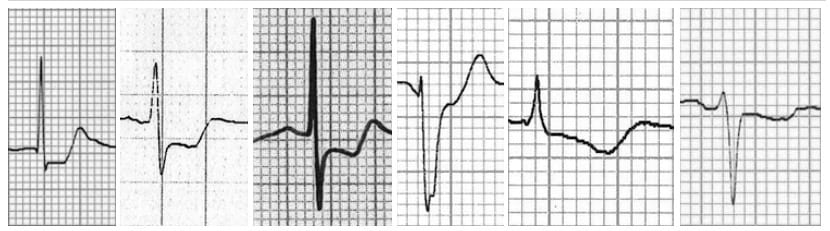
Identifying ischemic changes during ventricular activation and repolarization is one of the challenges physicians face when they are investigating a patient with pain in the chest or when they are trying to avoid ischemic events in the heart during an intervention.
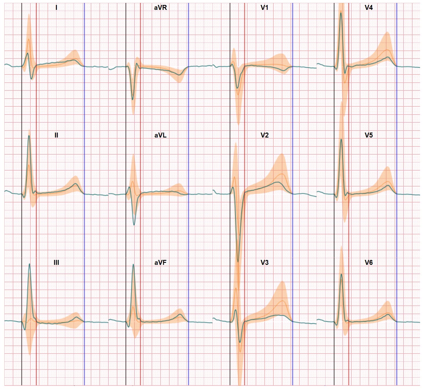
Using the 12 lead ECG for finding answers is standard practice but has severe limitations since the classic visualization of the ECG makes it hard to identify these ischemic events when the changes in the ST segment are hardly noticeable or do not occur (as far as the eye can see).
Improving the ECG interpretation on this challenge is a research topic which draws the attention of many cardiology researchers. Especially when they can use new techniques such as AI based data processing or additional visualisations of the inner workings of the heart as produced by Ultrasound or CT and MRI scans.
New options for early detection of ischemia in patients with inconclusive ECG patterns
Iris van der Schaaf is one of the brilliant young scientists who are working on this puzzle. And next week, during the annual conference of the International Society for Computerized Electrocardiology (ISCE), Iris is on of the 4 selected young investigators to present her work. Her presentation is part of the Jos Willems challenge on which we published an earlier blog. Iris study is focused on applying CineECG to support solving the Ischemia detection puzzle. And the results are so very promising!
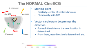
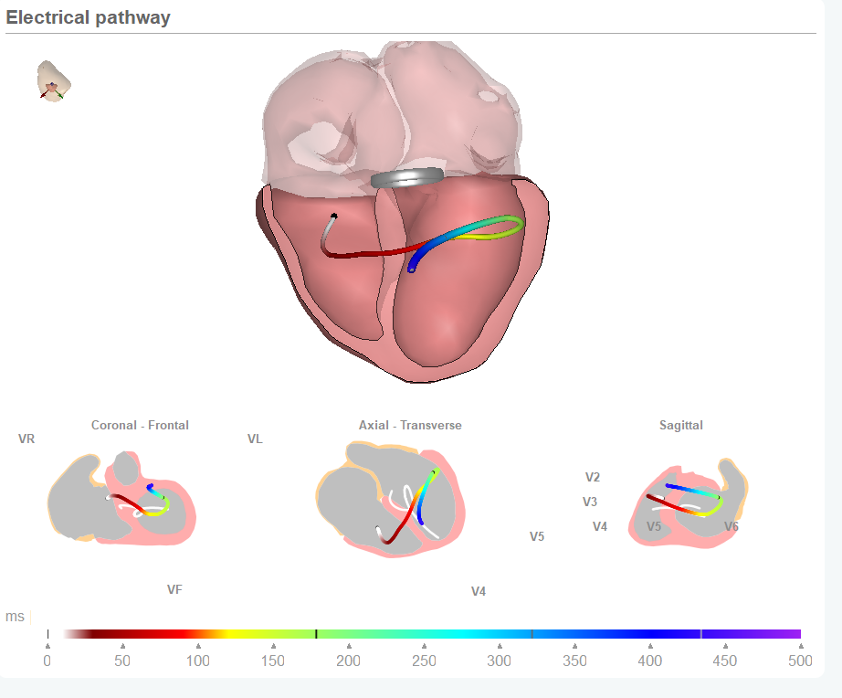
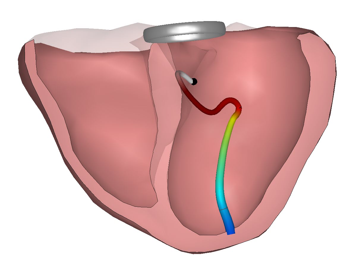
With CineECG the activation and repolarization pathway is visualized within the heart anatomy and this enables first to determine the electrical pathway of a normal activation. And within this normal pathway the direction and the location of the electrical pathway is determined for normal heart performance. This enters a new set of parameters based on the angle of the CIneECG at specific moments during the activation which are used by Iris in her search for early signs of Ischemia. Its true, you can visualize ischemic changes during ventricular activation!
Her project is still ongoing, but the initial findings show CIneECG is able to identify changes due to occlusions in all 3 coronary arteries (LAD, RCA, RCx) and that CineECG shows identifiable changes even when the ST segment changes are very small or no longer visible. And this truly great news since CineECG only requires 12 lead ECG data for input and the analysis is run in a split second.
We look forward to watching her presentation on March 31 2023 in Palm Springs California during ISCE 2023 and also look forward to the next steps with CIneECG in her research at UMCU and co-funded by the Dutch Heart Foundation.