In this blog we will address a case of identifying a specific case of a heart infarct, in particular an infarct in the lower part of the heart (Inferior wall infarction ; RCA distal to the RV branch). In an earlier blog we focused on an extensive anterior wall infarction and the ease of using CineECG to read the related ECG.
This distal RCA infarct happens when there’s a block or occlusion in a right coronary artery which supplies both the right ventricle but also the back side of the left ventricular septum, the wall separating the left and right chamber. In this example the infarct is located in this back left septal ventricular part of the heart. There are many places in the heart where a blockade can occur and knowing the location as soon as possible is important because the location is an indicator of the impact of the infarct. And thus the type and speed of therapy and intervention needed.
.
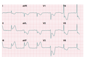
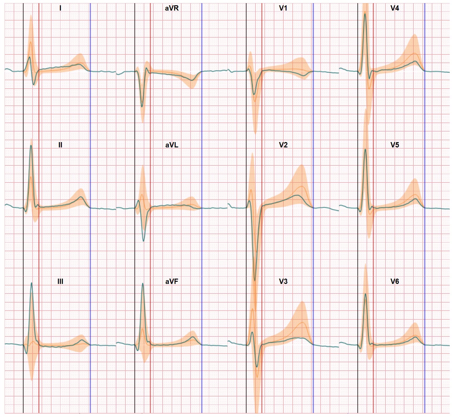
12-lead ECG data is first used to identify the location of the infarct and the way to do it is to look for specific patterns in the ECG graphs. If read correct, the ECG provides the distinct information from which a physician can make a decision and take action. There are many courses and instructions available to learn how to execute the ECG interpretation and for which ECG characteristics one should look (example here). However, not all ECG’s are interpreted correct or provide clear information. Of course a severe inferior infarct will be detected in most cases but when the deviations in the ECG are more subtle the assessment becomes more complicated. There are many courses and instructions available to learn how to execute the interpretation and for which ECG characteristics one should look. But in real life Physicians may decide to wait for more information (take another ECG at a later moment), use other diagnostics methods (blood test, X-ray, Echo, stress ECG, CRT) or take a closer look inside the artery (coronary angiogram). But all these other methods prolong the diagnostic time, increase the diagnostic costs and might create irreversible damage to the heart tissue.
CineECG supports effective detection of inferior wall infarction
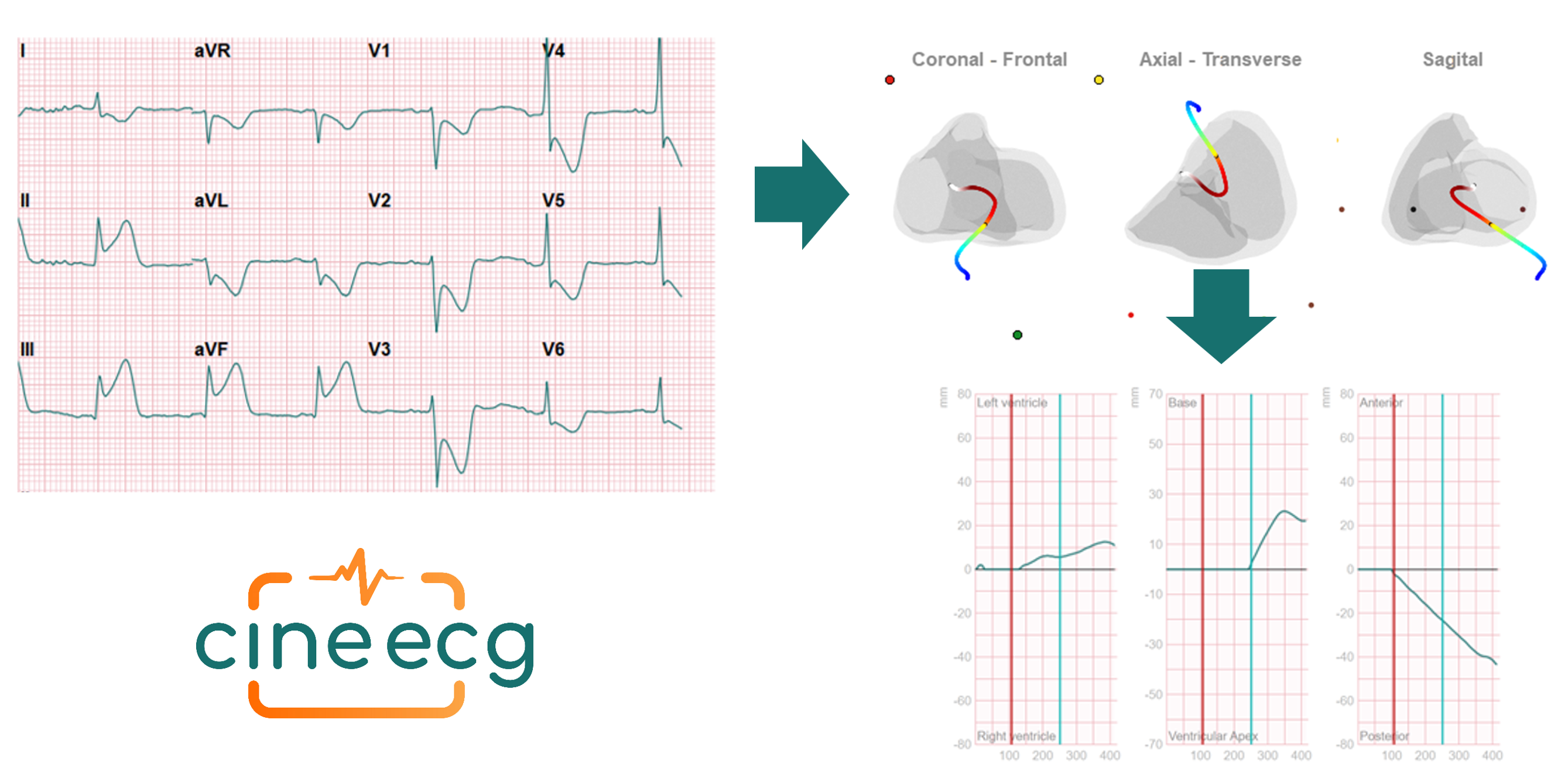
CineECG is a new method providing a new look at the same ECG and presents the data in a format which makes the ECG data easier to read and interpret[1]. CineECG shows the average path of the heart activation and recovery, visualizing the impact of blockades on this activation, supports the determination of the localisation of the blockade and compares the recorded data with normal healthy values. All to make the ECG assessment more accurate and reduce the need to use other diagnostics methods.
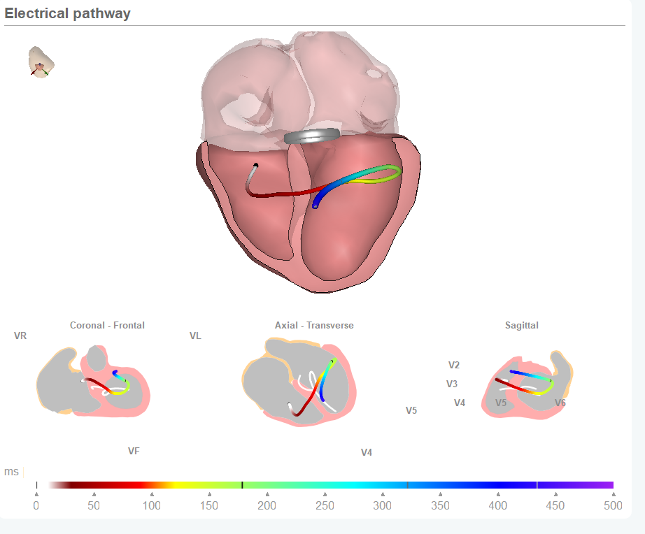
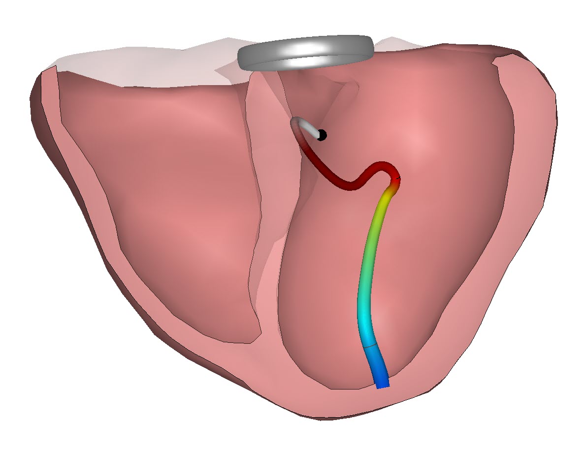
When applying CineECG in the case of an Inferior wall infarction; RCA distal of RV branch the analysis shows distinct deviations from a normal ECG. This is made visible by comparing the mean activation with the normal flow through the heart (shown as a white line in the 3D view / and shown as deviation from a horizontal line in the amplitude analysis. CineECG also indicates the position of the infarct both in the classic leads analysis and in the CineECG delta views. Plus the impact the infarct has on the repolarization (T waves deviate significant from normal and indicates the inferior location of the infarct).
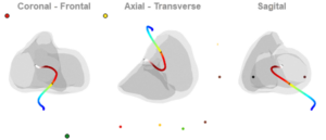
In more detail: in the coronal view (when we look straight at the heart as it was before us if we could look inside the torso) we see the T waves move to the front of the heart/body (blue line).
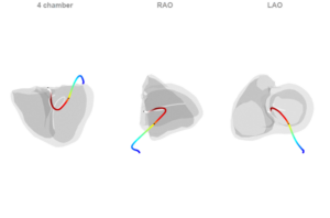
In the 4 chamber view you see the difference between the patient ECG and the normal CineECG which is shown as a white shadow. In the case of this inferior wall infarction the CineECG does not follow a normal pattern and the mean activation moves away from the apex, indicating the position of the obstruction in the inferior left wall.
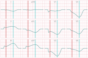
The two 2D delta graphs of the amplitudes and the movement of the CineECG through the heart support this finding. In these graphs CineECG shows also the deviations from normal. Both for the amplitudes and the mean activation path. And in this case we see ST elevations in leads II, III, and aVF as can be expected but also ST depression in leads I, VI-V6 which indicates an inferior infarct in the RCA.
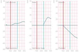
And we see a deviating T-wave direction in the inferior direction in the Anterior-Posterior Graph. And moving away from the apex Base-Apex view and moving towards the left ventricle in the LV-RV view , all indicating the location of the infarct.
This way using CineECG makes the identification of an inferior wall infarction easier and more specific.
[1] CineECG is a computational model system that analyses standard 12 lead ECGs and calculates
a range of quantitative parameters based on a mean representative QRS complex to the end of the T-wave. CineECG also produces a graphical 3D output that directly visualizes the temporo-spatial features of the cardiac activation and recovery, and integrates the electrical features with a
standardized cardiac anatomical model